Study: Overactive Immune Cells Linked to Common Form of Heart Failure
Tufts researchers identify T cell populations in mice with obesity and high blood pressure that infiltrate the heart, reducing the organ’s ability to pump blood
By Joseph Caputo
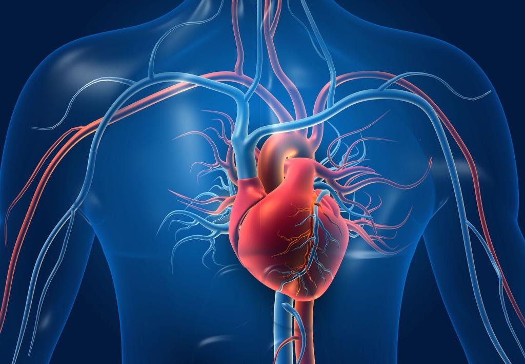
A patient admitted into the emergency room with shortness of breath and fatigue—concerning signs their heart may be struggling to pump blood—often presents with several comorbidities such as hypertension, obesity, and systemic chronic inflammation. In a Tufts-led study published October 24 in The Journal of Clinical Investigation, researchers explore the relationship between immune cell activity and heart failure, revealing unique T cell populations in mice with metabolic disorders that coincide with cardiac tissue abnormalities.
Using a recently developed mouse model that combines obesity and high blood pressure and which was developed at UT Southwestern Medical Center, the Tufts research team observed that these conditions yield the systemic activation of malfunctioning T cells that can stiffen the heart. When investigators removed these overactive immune populations, cardiac function was partially restored. The work raises the possibility that detecting or modulating cardiac inflammation in vulnerable patients could slow the progression of heart disease.
“Our paper is starting to tease apart what T cells originating from the periphery of the body are actually doing in the heart,” says first author Sasha Smolgovsky, a Ph.D. candidate in immunology at the Tufts Graduate School of Biomedical Sciences who was awarded two fellowships to pursue this work. “The heart is impacted almost as a secondary bystander, with the thickening of the tissue caused by inflammation leaving the heart unable to relax, thereby reducing cardiac output.”
This dysfunction, classified as heart failure with preserved ejection fraction (HFpEF), leaves the heart pumping blood but not relaxing enough to fill with a sufficient volume of blood. It is the slower-forming cousin of the better-known heart failure with reduced ejection fraction (HFrEF) typically seen in people who have just suffered a heart attack. HFpEF makes up about 50 percent of heart failure cases, but its pathology has been poorly understood, which Smolgovsky and colleagues are looking to address.
One of the study’s key findings is that the overactive T cells in the mouse model exhibited evidence of impaired resolution of cellular stress, which the research team want to explore as a potential clinical biomarker for HFpEF. They are also designing experiments to investigate how inflammation affects various types of heart cells to better characterize the role each plays in the disease.
“This story has unfolded in front of our eyes as the result of a hyper collaborative project that harnesses lessons from cancer inflammation, T cell biology and cardiology into something translational,” says senior author Pilar Alcaide, a Kenneth and JoAnn G. Wellner Professor in the Department of Immunology at the School of Medicine.
###
Smolgovsky, S., Bayer, A., Kaur, K., Sanders, E., Aronovitz, M., Filipp, M.E., Thorp, E.B., Schiattarella, G.G., Hill, J.A., Blanton, R.M., Cubillos-Ruiz, J.R., Alcaide, P. (2023). Impaired T cell IRE1α-XBP1 signaling directs inflammation in experimental Heart Failure with Preserved Ejection Fraction. Journal of Clinical Investigation. https://doi.org/10.1172/JCI171874.
Complete information on authors, funders, and conflicts of interest is available in the published paper.
DISCLAIMER: The content is solely the responsibility of the authors and does not necessarily represent the official views of the National Institutes of Health.
