Preclinical Imaging Core
The Tufts Preclinical Imaging Core (PIC) provides equipment and expertise for molecular and anatomical imaging for basic research and translational studies in live animals.
The Tufts Preclinical Imaging Core (PIC) provides equipment and expertise for molecular and anatomical imaging for basic research and translational studies in live animals.
The PIC not only supports research at Tufts University and affiliated hospitals, it also offers a wide range of contract research services for Biotech and Pharma.
Major instrumentation includes:
- PerkinElmer IVIS Spectrum CT Biophotonic Imager
- VisualSonics Vevo 2100 Ultrasound Imaging System
- Skyscan 1176 High Resolution Micro-CT Scanner
The technology provided by these instruments is applicable to a wide range of research areas that include tumor and stem cell biology, infectious disease, neurobiology, wound healing, and signal transduction. The PIC is also equipped to develop imageable animal models and perform surgical procedures and cell/tissue culture to support in vivo imaging and preclinical testing studies with these instruments.
For cancer researchers at academic institutions and companies, the PIC offers novel luciferase- and fluorescent protein-based syngeneic and xenograft models, as well as technical services for studying in vivo tumor growth and metastasis and preclinical drug testing. A technician is available for animal handling/husbandry, introduction of tumor cells and therapeutics into animals, and other services. Contact us for more information.
Instrument Use
Services provided by the Facility generally require technical assistance from Facility Staff. However, Tufts University and Tufts Medical Center users can be trained and certified to operate our instruments themselves at reduced ("Unassisted Use") cost.
|
Instrument Use (Charges per hour) |
Internal |
Outside Academic Institution |
Outside Commercial Institution |
|
PerkinElmer Spectrum CT Biophotonic Imager (charges per hour) |
$110.00 |
$143.00 |
$195.80 |
|
VisualSonics Vevo 2100 Ultrasound Imager (charges per hour) |
$110.00 |
$143.00 |
$195.80 |
|
Skyscan 1176 CT Scanner (charges per hour) |
$110.00 |
$143.00 |
$195.80 |
|
Iso anesthesia for live animal imaging (charges per hour) |
$34.00 |
$44.00 |
$60.00 |
Assistance with Imaging
|
Type of Assistance |
Internal |
Outside Academic Institution |
Outside Commercial Institution |
|
Facility Manager |
$90.00 |
$117.00 |
$160.20 |
|
Veterinarian/Veterinary Pathologist |
$163.00 |
$212.00 |
$290.00 |
|
Small Animal Imaging Analysis |
$112.00 |
$146.00 |
$200.00 |
Assistance with Projects
|
Projects |
Unassisted |
Assisted |
|
Custom Model Development |
n/a |
Inquire |
|
Drug Target Identification and Validation |
n/a |
Inquire |
|
Drug Testing (Efficacy, Safety, Biodistribution, and/or Pharmacokinetics) |
n/a |
Inquire |
|
Animal Pathology/Histology |
n/a |
Inquire |
|
In Vitro High Throughput Screening |
n/a |
Inquire |
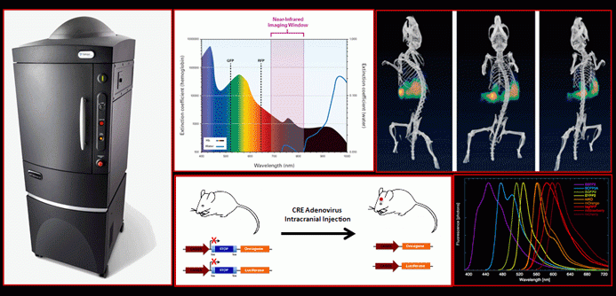
PerkinElmer IVIS Spectrum CT Biophotonic Imager
The PerkinElmer IVIS Spectrum CT Biophotonic Imager excels at imaging light emitted from small animals such as mice or rats but can also be used to image light emitted from Western blots, culture dishes and 96-well plates. The Spectrum CT can quantitate bioluminescent or chemiluminescent signals produced by luciferases and other reporters, as well as fluorescent signals produced by fluorescent proteins (GFP, RFP, etc.) and other fluorescent molecules. The instrument has the capability of imaging light in 2-D or pseudo 3-D mode and co-localizing light emission with anatomical features by micro-CT imaging.
SkyScan 1176 In Vivo Micro-CT
The SKYSCAN 1176 is a high performance preclinical in vivo micro-CT scanner. Its large format 11 megapixel x-ray camera gives high resolution and image field size at relatively rapid scan speeds. Image field width up to 68 mm allows full body mouse and rat scanning and distal limb scanning for big animals, such as rabbits, at voxel sizes of 9, 18 and 35µm. Variable x-ray applied voltage and filters provide scanning flexibility to allow imaging of a wide range of samples from lung tissue to bone. An integrated physiological monitoring system provides respiratory and cardiac gating for proven thoracic image improvement by synchronized acquisition.
VisualSonics Vevo 2100 Ultrasound Imager
The VisualSonics Vevo 770 Imager is a state of the art high frequency ultrasound imaging system designed for real-time imaging of internal soft tissues in mice and other small animals. The system is optimally configured for studies in the areas of cancer, vascular and stem cell biology. Microbubble contrast agents are available to facilitate blood flow and angiogenesis measurements and can be used to selectively deliver drugs to specific sites in the organism. The microinjection system that was purchased with the instrument allows image-guided injection or biopsy for introducing cells or other materials at specific organs sites or for removing tissue for biopsy.
General Services
- Instrumentation and expertise for molecular and anatomic imaging
- Full range of services for preclinical testing in animals including
- Model availability and development
- Animal care and handling
- Surgical procedures on small animals
- Administration of therapeutics
- Analysis of therapeutic safety and efficacy
- Pharmacokinetic and pharmacodynamic analyses
Cancer-related Services
- Model development specializing in imagable syngeneic tumor models administered orthotopically
- Availability of well characterized GEM, xenograft and syngeneic models for a wide range of cancers
- Drug target screening and validation
- Quantitative analysis of primary tumor growth and metastasis
- Full range of preclinical testing services for analysis of anti-cancer drugs and therapeutic regimens
- Analysis of therapeutic safety, efficacy
- Pharmacokinetics and pharmacodynamics
- Wide range of administration methods for tumors cells and therapeutics
- Immune and stromal cell profiling
- Image-guided injection and biopsy
Cancer Model Availability
Widely Used Luciferase-expressing Xenograft Models
- MDA-MB-231 (breast, ER-)
- MCF-7 (breast, ER+)
- BT-474 (breast, ER+, HER2+)
- MDA-MB-231 Human Breast Cancer Xenograft Model
- MCF-7 Human Breast Cancer Xenograft Model
- PC-3M (prostate)
- A549 (lung)
- HT-29 (colon)
- SKOV3 (ovarian)
- HeLa (cervical)
A wide range of other xenograft models are available on request.
Several Luciferase-expressing Syngeneic Models
- 4T1 metastatic breast
- 4T1 bone, brain, liver and lung tropic (metastatic breast)
- 66c14 (weakly metastatic breast)
- B16-F10 (melanoma)
- CT26 (colon)
- TRAMP (prostate)
- K8484, DT8082 (pancreatic)
Patient-derived Xenograft (PDX) Models
PDX models for breast cancer and several other cancers are available. These models, which are derived from well characterized patient tumors, involve expansion of tumor tissue in NOD/SCID/gamma (NSG) immunocompromised mice and subsequent implantation of tumor fragments in NSG mice for testing new drugs and therapeutic regimens. While PDX models, like other xenograft models, require the use of immunocompromised animals, they retain many of the characteristics of the tumors from which they were derived and provide a means of assessing the effects of therapeutics on specific cancer subtypes in a more physiological setting than can be achieved with conventional xenograft models.